251:
239:
218:-secreting cells. The thick middle layer consists of interspersed elastin fibers, collagen bundles, and fibroblasts. The most superficial layer is made up of loose connective tissue which contains vascular channels. Combined these three layers create a smooth gliding surface as well as mechanically strong tissue which prevents tendon bowstringing. The extensor retinaculum of the foot has similar structure.
29:
190:
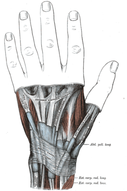
There are six separate synovial sheaths run beneath the extensor retinaculum: (1st) abductor pollicis longus and extensor pollicis brevis tendons, (2nd) extensor carpi radialis longus and brevis tendons, (3rd) extensor pollicis longus tendon, (4th) extensor digitorium communis and extensor indicis
226:
Studies conducted on the retinaculum have exhibited it to have several possible surgical treatments uses. A graft of the extensor retinaculum was shown to be useful in treating boxer's knuckle when direct repair of the damaged capsule is not possible. Because of their similarities in histological
198:
corresponds in location and structure to the extensor retinaculum, both being formations of the antebrachial fascia and therefore continuous. Consequently, the
98:
580:
359:
382:. Friedrich Paulsen, Tobias M. Böckers, J. Waschke, Stephan Winkler, Katja Dalkowski, Jörg Mair, Sonja Klebe, Elsevier ClinicalKey. Munich. 2019.
227:
structure, studies also show the extensor retinaculum to be a reasonable biological replacement for reconstruction of a deficient annular pulley.
363:
411:
74:
1178:
573:
1183:
972:
968:
895:
438:
387:
238:
939:
856:
787:
143:
792:
566:
250:
170:
The extensor retinaculum is a strong, fibrous band, extending obliquely downward and medialward across the back of the
920:
335:
731:
1145:
1029:
1024:
1019:
846:
703:
1277:
199:
1216:
1173:
1140:
989:
984:
925:
93:
553:
454:
Klein, David M.; Katzman, Barry M.; Mesa, Joseph A.; Lipton, Jeffrey F.; Caligiuri, Daniel A. (1999-01-01).
330:. Vogl, Wayne., Mitchell, Adam W. M., Gray, Henry, 1825-1861. Philadelphia: Elsevier/Churchill Livingstone.
885:
214:
Structurally, the retinaculum consists of three layers. The deepest layer, the gliding layer, consists of
890:
693:
415:
1221:
979:
915:
749:
505:
455:
275:
1152:
1135:
1036:
711:
1311:
1206:
1190:
1068:
880:
739:
195:
191:
proprius tendons, (5th) extensor digiti minimi tendon and (6th) extensor carpi ulnaris tendon.
159:
105:
81:
69:
1101:
1058:
875:
151:
1073:
638:
628:
623:
8:
1168:
1080:
1014:
825:
674:
669:
135:
1282:
1257:
815:
645:
633:
405:
353:
303:
291:
721:
533:
525:
483:
475:
434:
393:
383:
341:
331:
295:
307:
1009:
820:
517:
467:
287:
1130:
963:
777:
772:
767:
744:
215:
1096:
664:
613:
521:
504:
Nagaoka, Masahiro; Satoh, Takako; Nagao, Soya; Matsuzaki, Hiromi (2006-07-01).
397:
276:"The Extensor Retinaculum of The Wrist: An Anatomical and Biomechanical Study"
1305:
529:
479:
86:
345:
16:
A thickened fascia holding the tendons of the hand extensor muscles in place
618:
537:
487:
471:
377:
299:
274:
PALMER, A. K.; SKAHEN, J. R.; WERNER, F. W.; GLISSON, R. R. (2016-11-17).
325:
175:
111:
958:
323:
558:
601:
456:"Histology of the Extensor Retinaculum of the Wrist and the Ankle"
841:
179:
147:
34:
1233:
1050:
759:
716:
655:
589:
139:
429:
Moore, Keith L.; Dalley, Arthur F.; Agur, Anne M. R. (2018).
171:
57:
38:
1115:
554:
Hand kinesiology at the
University of Kansas Medical Center
155:
503:
182:, strengthened by the addition of some transverse fibers.
28:
688:
593:
273:
244:
Transverse section across distal ends of radius and ulna.
506:"Extensor Retinaculum Graft for Chronic Boxer's Knuckle"
162:(which is located on the anterior side of the forearm).
453:
41:. (Dorsal carpal ligament labeled at bottom center.)
256:Extensor retinaculum of the hand. Deep dissection.
1303:
428:
324:Drake, Richard L. (Richard Lee), 1950- (2005).
574:
433:(8th ed.). Wolters Kluwer. p. 159.
358:: CS1 maint: multiple names: authors list (
146:in place. It is located on the back of the
581:
567:
410:: CS1 maint: location missing publisher (
362:) CS1 maint: numeric names: authors list (
27:
221:
63:retinaculum musculorum extensorum manus
1304:
588:
562:
499:
497:
319:
317:
230:
194:On the dorsal side of the hand, the
13:
422:
14:
1323:
547:
494:
447:
314:
267:
249:
237:
134:) is a thickened portion of the
22:Extensor retinaculum of the hand
202:is commonly referred to as the
969:extensor carpi radialis longus
896:flexor digitorum superficialis
370:
1:
292:10.1016/S0266-7681(85)80006-1
261:
174:. It consists of part of the
209:
185:
165:
158:. It is continuous with the
7:
1179:flexor digiti minimi brevis
510:The Journal of Hand Surgery
460:The Journal of Hand Surgery
431:Clinically Oriented Anatomy
327:Gray's anatomy for students
10:
1328:
921:flexor digitorum profundus
522:10.1016/j.jhsa.2006.02.027
204:transverse carpal ligament
132:posterior annular ligament
33:The mucous sheaths of the
1266:
1241:
1232:
1199:
1161:
1123:
1114:
1089:
1049:
998:
947:
938:
904:
864:
855:
840:
803:
758:
730:
702:
687:
654:
609:
600:
104:
92:
80:
68:
56:
51:
46:
26:
21:
1146:abductor pollicis brevis
1030:extensor pollicis longus
1025:extensor pollicis brevis
1020:abductor pollicis longus
379:Sobotta anatomy textbook
280:Journal of Hand Surgery
1184:abductor digiti minimi
1174:opponens digiti minimi
1141:flexor pollicis brevis
990:extensor carpi ulnaris
985:extensor digiti minimi
926:flexor pollicis longus
472:10.1053/jhsu.1999.0799
196:palmar carpal ligament
160:palmar carpal ligament
128:dorsal carpal ligament
106:Anatomical terminology
1059:bicipital aponeurosis
886:flexor carpi radialis
414:) CS1 maint: others (
222:Clinical significance
1253:extensor retinaculum
891:flexor carpi ulnaris
206:to avoid confusion.
124:extensor retinaculum
1081:antebrachial fascia
1015:anatomical snuffbox
826:triangular interval
783:intermuscular septa
675:infraspinous fascia
670:supraspinous fascia
178:of the back of the
136:antebrachial fascia
37:on the back of the
1283:palmar aponeurosis
1278:flexor retinaculum
1258:extensor expansion
1102:osborne's ligament
980:extensor digitorum
916:pronator quadratus
816:quadrangular space
750:articularis cubiti
200:flexor retinaculum
1299:
1298:
1295:
1294:
1291:
1290:
1153:adductor pollicis
1136:opponens pollicis
1110:
1109:
1045:
1044:
934:
933:
836:
835:
683:
682:
440:978-1-4963-4721-3
389:978-0-7020-6760-0
231:Additional images
120:
119:
115:
1319:
1271:
1246:
1239:
1238:
1121:
1120:
1037:extensor indicis
1003:
952:
945:
944:
909:
869:
862:
861:
853:
852:
821:triangular space
712:coracobrachialis
700:
699:
607:
606:
583:
576:
569:
560:
559:
542:
541:
501:
492:
491:
451:
445:
444:
426:
420:
419:
409:
401:
374:
368:
367:
357:
349:
321:
312:
311:
271:
253:
241:
144:extensor muscles
112:edit on Wikidata
109:
31:
19:
18:
1327:
1326:
1322:
1321:
1320:
1318:
1317:
1316:
1302:
1301:
1300:
1287:
1267:
1262:
1242:
1228:
1195:
1191:palmaris brevis
1157:
1106:
1085:
1041:
999:
994:
964:brachioradialis
948:
930:
905:
900:
881:palmaris longus
865:
844:
832:
799:
778:brachial fascia
773:axillary fascia
768:axillary sheath
754:
740:triceps brachii
726:
691:
679:
650:
596:
587:
550:
545:
502:
495:
452:
448:
441:
427:
423:
403:
402:
390:
376:
375:
371:
351:
350:
338:
322:
315:
272:
268:
264:
257:
254:
245:
242:
233:
224:
216:hyaluronic acid
212:
188:
168:
138:that holds the
116:
42:
17:
12:
11:
5:
1325:
1315:
1314:
1297:
1296:
1293:
1292:
1289:
1288:
1286:
1285:
1280:
1274:
1272:
1264:
1263:
1261:
1260:
1255:
1249:
1247:
1236:
1230:
1229:
1227:
1226:
1225:
1224:
1219:
1209:
1203:
1201:
1197:
1196:
1194:
1193:
1188:
1187:
1186:
1181:
1176:
1165:
1163:
1159:
1158:
1156:
1155:
1150:
1149:
1148:
1143:
1138:
1127:
1125:
1118:
1112:
1111:
1108:
1107:
1105:
1104:
1099:
1097:cubital tunnel
1093:
1091:
1087:
1086:
1084:
1083:
1078:
1077:
1076:
1071:
1064:common tendons
1061:
1055:
1053:
1047:
1046:
1043:
1042:
1040:
1039:
1034:
1033:
1032:
1027:
1022:
1012:
1006:
1004:
996:
995:
993:
992:
987:
982:
977:
976:
975:
966:
955:
953:
942:
936:
935:
932:
931:
929:
928:
923:
918:
912:
910:
902:
901:
899:
898:
893:
888:
883:
878:
876:pronator teres
872:
870:
859:
850:
838:
837:
834:
833:
831:
830:
829:
828:
823:
818:
807:
805:
801:
800:
798:
797:
796:
795:
790:
780:
775:
770:
764:
762:
756:
755:
753:
752:
747:
742:
736:
734:
728:
727:
725:
724:
719:
714:
708:
706:
697:
685:
684:
681:
680:
678:
677:
672:
667:
665:deltoid fascia
661:
659:
652:
651:
649:
648:
643:
642:
641:
636:
631:
626:
616:
610:
604:
598:
597:
586:
585:
578:
571:
563:
557:
556:
549:
548:External links
546:
544:
543:
516:(6): 947–951.
493:
466:(4): 799–802.
446:
439:
421:
388:
369:
336:
313:
265:
263:
260:
259:
258:
255:
248:
246:
243:
236:
232:
229:
223:
220:
211:
208:
187:
184:
167:
164:
118:
117:
108:
102:
101:
96:
90:
89:
84:
78:
77:
72:
66:
65:
60:
54:
53:
49:
48:
44:
43:
32:
24:
23:
15:
9:
6:
4:
3:
2:
1324:
1313:
1312:Human anatomy
1310:
1309:
1307:
1284:
1281:
1279:
1276:
1275:
1273:
1270:
1265:
1259:
1256:
1254:
1251:
1250:
1248:
1245:
1240:
1237:
1235:
1231:
1223:
1220:
1218:
1215:
1214:
1213:
1210:
1208:
1205:
1204:
1202:
1198:
1192:
1189:
1185:
1182:
1180:
1177:
1175:
1172:
1171:
1170:
1167:
1166:
1164:
1160:
1154:
1151:
1147:
1144:
1142:
1139:
1137:
1134:
1133:
1132:
1129:
1128:
1126:
1124:lateral volar
1122:
1119:
1117:
1113:
1103:
1100:
1098:
1095:
1094:
1092:
1088:
1082:
1079:
1075:
1072:
1070:
1067:
1066:
1065:
1062:
1060:
1057:
1056:
1054:
1052:
1048:
1038:
1035:
1031:
1028:
1026:
1023:
1021:
1018:
1017:
1016:
1013:
1011:
1008:
1007:
1005:
1002:
997:
991:
988:
986:
983:
981:
978:
974:
970:
967:
965:
962:
961:
960:
957:
956:
954:
951:
946:
943:
941:
937:
927:
924:
922:
919:
917:
914:
913:
911:
908:
903:
897:
894:
892:
889:
887:
884:
882:
879:
877:
874:
873:
871:
868:
863:
860:
858:
854:
851:
848:
843:
839:
827:
824:
822:
819:
817:
814:
813:
812:
809:
808:
806:
802:
794:
791:
789:
786:
785:
784:
781:
779:
776:
774:
771:
769:
766:
765:
763:
761:
757:
751:
748:
746:
743:
741:
738:
737:
735:
733:
729:
723:
720:
718:
715:
713:
710:
709:
707:
705:
701:
698:
695:
690:
686:
676:
673:
671:
668:
666:
663:
662:
660:
657:
653:
647:
644:
640:
639:subscapularis
637:
635:
632:
630:
629:infraspinatus
627:
625:
624:supraspinatus
622:
621:
620:
617:
615:
612:
611:
608:
605:
603:
599:
595:
591:
584:
579:
577:
572:
570:
565:
564:
561:
555:
552:
551:
539:
535:
531:
527:
523:
519:
515:
511:
507:
500:
498:
489:
485:
481:
477:
473:
469:
465:
461:
457:
450:
442:
436:
432:
425:
417:
413:
407:
399:
395:
391:
385:
381:
380:
373:
365:
361:
355:
347:
343:
339:
337:0-443-06612-4
333:
329:
328:
320:
318:
309:
305:
301:
297:
293:
289:
285:
281:
277:
270:
266:
252:
247:
240:
235:
234:
228:
219:
217:
207:
205:
201:
197:
192:
183:
181:
177:
173:
163:
161:
157:
153:
149:
145:
141:
137:
133:
129:
125:
113:
107:
103:
100:
97:
95:
91:
88:
85:
83:
79:
76:
73:
71:
67:
64:
61:
59:
55:
50:
45:
40:
36:
30:
25:
20:
1268:
1252:
1243:
1211:
1200:intermediate
1162:medial volar
1063:
1000:
950:superficial:
949:
906:
867:superficial:
866:
847:compartments
810:
782:
694:compartments
619:rotator cuff
513:
509:
463:
459:
449:
430:
424:
378:
372:
326:
286:(1): 11–16.
283:
279:
269:
225:
213:
203:
193:
189:
169:
131:
127:
123:
121:
75:A04.6.03.010
62:
646:teres major
634:teres minor
176:deep fascia
52:Identifiers
1244:posterior:
1212:interossei
1169:hypothenar
959:mobile wad
722:brachialis
398:1132300315
262:References
1269:anterior:
1207:lumbrical
1010:supinator
940:posterior
732:posterior
530:0363-5023
480:0363-5023
406:cite book
354:cite book
210:Histology
186:Relations
166:Structure
1306:Category
1069:extensor
857:anterior
745:anconeus
704:anterior
602:Shoulder
538:16843154
488:10447172
346:55139039
308:11507945
152:proximal
842:Forearm
788:lateral
614:deltoid
592:of the
590:Muscles
300:3998587
180:forearm
154:to the
150:, just
148:forearm
142:of the
140:tendons
47:Details
35:tendons
1234:fascia
1222:palmar
1217:dorsal
1131:thenar
1074:flexor
1051:fascia
973:brevis
811:spaces
793:medial
760:fascia
717:biceps
656:fascia
536:
528:
486:
478:
437:
396:
386:
344:
334:
306:
298:
1090:other
1001:deep:
907:deep:
804:other
304:S2CID
172:wrist
130:, or
110:[
99:39987
58:Latin
39:wrist
1116:Hand
971:and
534:PMID
526:ISSN
484:PMID
476:ISSN
435:ISBN
416:link
412:link
394:OCLC
384:ISBN
364:link
360:link
342:OCLC
332:ISBN
296:PMID
156:hand
122:The
87:2546
70:TA98
689:Arm
594:arm
518:doi
468:doi
288:doi
94:FMA
82:TA2
1308::
532:.
524:.
514:31
512:.
508:.
496:^
482:.
474:.
464:24
462:.
458:.
408:}}
404:{{
392:.
356:}}
352:{{
340:.
316:^
302:.
294:.
284:10
282:.
278:.
849:)
845:(
696:)
692:(
658::
582:e
575:t
568:v
540:.
520::
490:.
470::
443:.
418:)
400:.
366:)
348:.
310:.
290::
126:(
114:]
Text is available under the Creative Commons Attribution-ShareAlike License. Additional terms may apply.


